Ureterocele
Incontinence - ureterocele
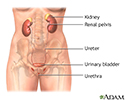
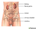
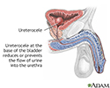
A ureterocele is a swelling at the bottom of one of the ureters. Ureters are the tubes that carry urine from the kidney to the bladder. The swollen area can block urine flow.
A ureterocele is a birth defect.
Causes
A ureterocele occurs in the lower part of the ureter, where it enters the bladder. The swollen area prevents urine from moving freely into the bladder. The urine collects in the ureterocele and stretches its walls. It expands like a water balloon.
A ureterocele can also cause urine to flow backward from the bladder to the kidney. This is called reflux which may damage the kidney.
Ureteroceles occur in about 1 in 500 people. This condition is equally common in both the left and right ureters.
Symptoms
Most people with ureteroceles do not have any symptoms. When symptoms do occur, they may include:
- Abdominal pain
- Back pain that may be only on one side
- Severe side (flank) pain and spasms that may reach to the groin, genitals, and thigh
- Blood in the urine
- Burning pain while urinating (dysuria)
- Fever
- Difficulty starting urine flow or slowing of urine flow
- Urinary tract infection
Some other symptoms are:
- Foul-smelling urine
- Frequent and urgent urination
- Lump (mass) in the abdomen that can be felt
- Ureterocele tissue falls down (prolapse) through the female urethra and into the vagina
- Urinary incontinence
Exams and Tests
Large ureteroceles are often diagnosed earlier than smaller ones. Ureteroceles are often discovered by a pregnancy-related ultrasound before the baby is born.
Some people with ureteroceles do not know they have the condition. Often, the problem is found later in life due to kidney stones or infection.
A urinalysis may reveal blood in the urine or signs of urinary tract infection.
The following tests may be done:
- Abdominal ultrasound
- CT scan of the abdomen
- Cystoscopy (examination of the inside of the bladder)
- Pyelogram (retrograde)
- Radionuclide renal scan
- Voiding cystourethrogram
Blood pressure may be high if there is kidney damage.
Treatment
Antibiotics are often given to prevent further infections until surgery can be done.
The goal of treatment is to eliminate the blockage. Drains placed in the ureter or renal area (stents) may provide short-term relief of symptoms.
Surgery to repair the ureterocele cures the condition in most cases. Your surgeon may cut into the ureterocele. Another surgery may involve removing the ureterocele and reattaching the ureter to the bladder. The type of surgery depends on your age, overall health, and extent of the blockage.
Outlook (Prognosis)
The outcome varies. The damage may be temporary if the blockage can be cured. However, damage to the kidney may be permanent if the condition doesn't go away.
Kidney failure is uncommon. The other kidney will most often work normally.
Possible Complications
Complications may include:
- Long-term bladder damage (urinary retention)
- Long-term kidney damage, including loss of function in one kidney
- Urinary tract infection that keeps coming back
When to Contact a Medical Professional
Contact your health care provider if you have symptoms of ureterocele.
References
Guay-Woodford LM. Hereditary nephropathies and developmental renal/urinary abnormalities. In: Goldman L, Cooney KA, eds. Goldman-Cecil Medicine. 27th ed. Philadelphia, PA: Elsevier; 2024:chap 113.
Stanasel I, Peters CA. Ectopic ureter, ureterocele, and ureteral anomalies. In: Partin AW, Dmochowski RR, Kavoussi LR, Peters CA, eds. Campbell-Walsh-Wein Urology. 12th ed. Philadelphia, PA: Elsevier; 2021:chap 41.
Review Date: 9/2/2024


