
Uvea
Vascular tunic
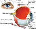
The uvea is the middle layer of the eye. It lies beneath the white part of the eye (the sclera). It is made of the iris, ciliary body, and choroid. These structures control many eye functions, including adjusting to different levels of light or distances of objects. Inflammation of one or more of these structures is called uveitis.


References
Evans M. Anatomy of the uvea. In: Yanoff M, Duker JS, eds. Ophthalmology. 6th ed. Philadelphia, PA: Elsevier; 2023:chap 7.1.
Taber's Cyclopedic Medical Dictionary. 24th ed. F.A. Davis Company; 2021. www.tabers.com/tabersonline/view/Tabers-Dictionary/770909/0/uvea?q=uvea. Accessed November 3, 2023.
Review Date: 11/1/2023
Reviewed By: Linda J. Vorvick, MD, Clinical Professor, Department of Family Medicine, UW Medicine, School of Medicine, University of Washington, Seattle, WA. Also reviewed by David C. Dugdale, MD, Medical Director, Brenda Conaway, Editorial Director, and the A.D.A.M. Editorial team.
 All rights reserved.