Lima Memorial Health System Logo
Approximate ER WAIT TIME
5

Health Library

Uvea
     
Print-Friendly
Bookmarks

Uvea

Vascular tunic


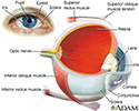
The uvea is the middle layer of the eye. It lies beneath the white part of the eye (the sclera). It is made of the iris, ciliary body, and choroid. These structures control many eye functions, including adjusting to different levels of light or distances of objects. Inflammation of one or more of these structures is called uveitis.


References

Evans M. Anatomy of the uvea. In: Yanoff M, Duker JS, eds. Ophthalmology. 6th ed. Philadelphia, PA: Elsevier; 2023:chap 7.1.

Taber's Cyclopedic Medical Dictionary. 24th ed. F.A. Davis Company; 2021. www.tabers.com/tabersonline/view/Tabers-Dictionary/770909/0/uvea?q=uvea. Accessed November 3, 2023.

BACK TO TOPText only

 
  • Eye

    Eye

    illustration

    • Eye

      Eye

      illustration

    Tests for Uvea


      Review Date: 11/1/2023

      Reviewed By: Linda J. Vorvick, MD, Clinical Professor, Department of Family Medicine, UW Medicine, School of Medicine, University of Washington, Seattle, WA. Also reviewed by David C. Dugdale, MD, Medical Director, Brenda Conaway, Editorial Director, and the A.D.A.M. Editorial team.

      The information provided herein should not be used during any medical emergency or for the diagnosis or treatment of any medical condition. A licensed medical professional should be consulted for diagnosis and treatment of any and all medical conditions. Links to other sites are provided for information only -- they do not constitute endorsements of those other sites. No warranty of any kind, either expressed or implied, is made as to the accuracy, reliability, timeliness, or correctness of any translations made by a third-party service of the information provided herein into any other language. © 1997- A.D.A.M., a business unit of Ebix, Inc. Any duplication or distribution of the information contained herein is strictly prohibited.
      © 1997- adam.comAll rights reserved.