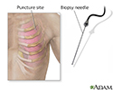
Pleural needle biopsy
Closed pleural biopsy; Needle biopsy of the pleura
Pleural biopsy is a procedure to remove a sample of the pleura. This is the thin tissue that lines the chest cavity and surrounds the lungs. The biopsy is done to check the pleura for disease or infection.
How the Test is Performed
This test may be done in the hospital. It may also be done at a clinic or health care provider's office.
The procedure involves the following:
- You will be sitting up during the procedure.
- Your provider cleanses the skin at the biopsy site.
- A numbing drug (anesthetic) is injected through the skin and into the lining of the lungs and chest wall (pleural membrane).
- A larger, hollow needle is then placed gently through the skin into the chest cavity. Sometimes, your provider uses ultrasound or CT imaging to guide the needle.
- A smaller cutting needle inside the hollow one is used to collect tissue samples. During this part of the procedure, you are asked to sing, hum, or say "eee." This helps prevent air from getting into the chest cavity, which can cause the lung to collapse (pneumothorax). Usually, 3 or more biopsy samples are taken.
- When the test is finished, a bandage is placed over the biopsy site.
In recent years, pleural biopsy is most often done using a fiberoptic scope. The scope allows your provider to view the area of the pleura from which the biopsies are taken.
How to Prepare for the Test
You will have blood tests before the biopsy. You will likely have a chest x-ray.
How the Test will Feel
When the local anesthetic is injected, you may feel a brief prick and a burning sensation. When the biopsy needle is inserted, you may feel pressure. As the needle is being removed, you may feel tugging.
Why the Test is Performed
Pleural biopsy is often done to find the cause of a collection of fluid around the lung (pleural effusion) or other abnormality of the pleural membrane. Pleural biopsy can diagnose tuberculosis, cancer, and other diseases.
If this type of pleural biopsy is not enough to make a diagnosis, you may need a surgical biopsy of the pleura.
Normal Results
Pleural tissues appear normal, without signs of inflammation, infection, or cancer.
What Abnormal Results Mean
Abnormal results may reveal:
- Primary lung cancer
- Malignant mesothelioma
- Metastatic pleural tumor
- Collagen vascular disease
- Tuberculosis
- Other infections
Risks
There is a slight chance of the needle puncturing the wall of the lung, which can partially collapse the lung. This usually gets better on its own. Sometimes, a chest tube is needed to drain the air and expand the lung.
There is also a chance of excessive blood loss.
Considerations
If a closed pleural biopsy is not enough to make a diagnosis, you may need a surgical biopsy of the pleura. This procedure has been mostly replaced by a procedure that uses a scope to visualize the pleura while taking the biopsy (pleuroscopy).
References
Reed JC. Pleural effusions. In: Reed JC, ed. Chest Radiology: Patterns and Differential Diagnoses. 7th ed. Philadelphia, PA: Elsevier; 2018:chap 4.
Walsh R, Klein JS. Thoracic radiology: invasive diagnostic imaging and image-guided interventions. In: Broaddus VC, Ernst JD, King TE, et al, eds. Murray and Nadel's Textbook of Respiratory Medicine. 7th ed. Philadelphia, PA: Elsevier; 2022:chap 21.
Review Date: 8/13/2023
Reviewed By: Denis Hadjiliadis, MD, MHS, Paul F. Harron Jr. Professor of Medicine, Pulmonary, Allergy, and Critical Care, Perelman School of Medicine, University of Pennsylvania, Philadelphia, PA. Also reviewed by David C. Dugdale, MD, Medical Director, Brenda Conaway, Editorial Director, and the A.D.A.M. Editorial team.


