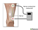
Nerve conduction velocity
NCV; Nerve conduction study (NCS)
Nerve conduction velocity (NCV) is a test to see how fast electrical signals move through a nerve. This test is done along with electromyography (EMG) to assess the muscles for abnormalities.
How the Test is Performed
Adhesive patches called surface electrodes are placed on the skin over nerves or muscles at different spots. A very mild electrical impulse is applied via other patches or a handheld stimulator to stimulate the nerve.
The resulting electrical activity of the nerve is recorded by the electrodes. The distance between electrodes and the time it takes for electrical impulses to travel between electrodes are used to measure the speed of the nerve signals. It is normal for several different nerves to be tested.
EMG is the recording from needles placed into the muscles. This is often done during the same visit as NCV.
How to Prepare for the Test
You must stay at a normal body temperature. Being too cold alters nerve conduction and can give false results.
Tell your health care provider if you have a cardiac defibrillator or pacemaker, or other implanted device such as a deep brain stimulator. Special steps may need to be taken before the test if you have one of these devices.
Do not wear any lotions, sunscreen, perfume, or moisturizer on your body on the day of the test.
How the Test will Feel
The impulse may feel like an electric shock. You may feel some discomfort depending on how strong the impulse is. You should feel no pain once the test is finished.
Often, the nerve conduction test is followed by EMG. In this test, a needle is placed into a muscle and you are told to contract that muscle. This process can be uncomfortable during the test. You may have muscle soreness or bruising after the test at the site where the needle was inserted.
Why the Test is Performed
This test is used to diagnose nerve damage or destruction. The test may sometimes be used to evaluate diseases of nerve or muscle, including:
- Myopathy
- Lambert-Eaton syndrome
- Myasthenia gravis
- Carpal tunnel syndrome
- Tarsal tunnel syndrome
- Diabetic neuropathy
- Bell palsy
- Guillain-Barré syndrome
- Brachial plexopathy
Normal Results
NCV is related to the diameter of the nerve and the degree of myelination (the presence of a myelin sheath on the nerve fiber) of the nerve. Newborn infants have values that are approximately half that of adults. Adult values are normally reached by age 3 or 4.
Note: Normal value ranges may vary slightly among different laboratories. Talk to your provider about the meaning of your specific test results.
What Abnormal Results Mean
Most often, abnormal results are due to nerve damage or destruction, including:
- Axonopathy (damage to the long portion of the nerve cell)
- Conduction block (the impulse is blocked somewhere along the nerve pathway)
- Demyelination (damage and loss of the fatty insulation surrounding the nerve cell)
The nerve damage or destruction may be due to many different conditions, including:
- Alcoholic neuropathy
- Diabetic neuropathy
- Nerve effects of uremia (from kidney failure)
- Traumatic injury to a nerve
- Guillain-Barré syndrome
- Diphtheria
- Carpal tunnel syndrome
- Brachial plexopathy
- Charcot-Marie-Tooth disease (hereditary)
- Chronic inflammatory polyneuropathy
- Common peroneal/fibular nerve dysfunction
- Distal median nerve dysfunction
- Femoral nerve dysfunction
- Friedreich ataxia
- Mononeuritis multiplex (multiple mononeuropathies)
- Primary amyloidosis
- Radial nerve dysfunction
- Sciatic nerve dysfunction
- Secondary systemic amyloidosis
- Sensorimotor polyneuropathy
- Tibial nerve dysfunction
- Ulnar nerve dysfunction
Any peripheral neuropathy can cause abnormal results. Damage to the spinal cord and disk herniation (herniated nucleus pulposus) with nerve root compression can also cause abnormal results.
Considerations
An NCV test shows the condition of the best surviving nerve fibers. Therefore, in some cases the results may be normal, even if there is nerve damage.
References
Deluca GC, Griggs RC. Approach to the patient with neurologic disease. In: Goldman L, Schafer AI, eds. Goldman-Cecil Medicine. 26th ed. Philadelphia, PA: Elsevier; 2020:chap 368.
Nuwer MR, Pouratian N. Monitoring of neural function: electromyography, nerve conduction, and evoked potentials. In: Winn HR, ed. Youmans and Winn Neurological Surgery. 8th ed. Philadelphia, PA: Elsevier; 2023:chap 274.
Review Date: 4/29/2023
Reviewed By: Joseph V. Campellone, MD, Department of Neurology, Cooper Medical School of Rowan University, Camden, NJ. Review provided by VeriMed Healthcare Network. Also reviewed by David C. Dugdale, MD, Medical Director, Brenda Conaway, Editorial Director, and the A.D.A.M. Editorial team.


